Digital and telepathology platform
that empowers pathologists
Digital and telepathology platform trusted by
LIS for Pathologists
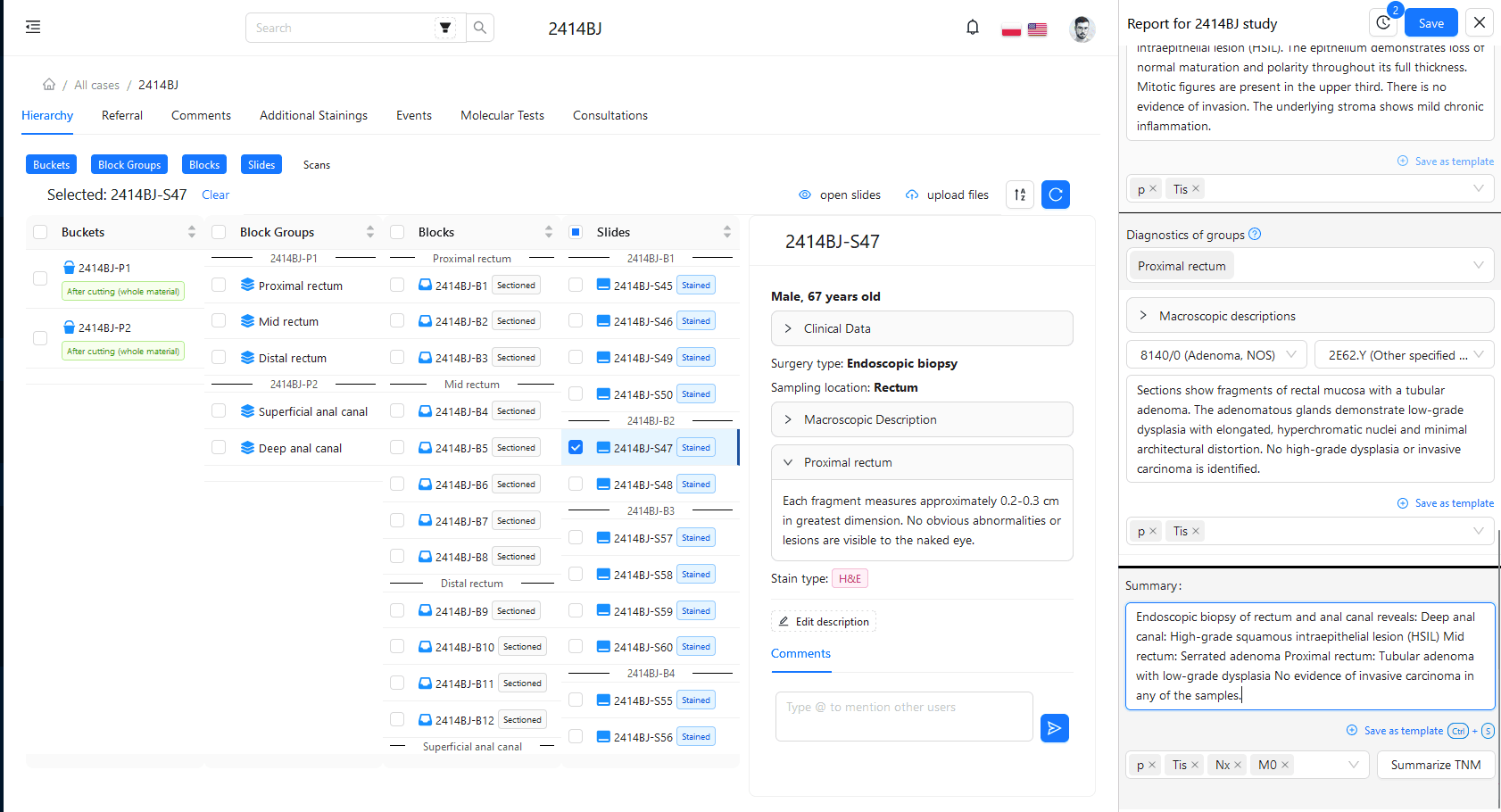
Streamline your laboratory operations with our comprehensive LIMS module. Efficiently manage test registration, process tracking, and result reporting. The system allows full control over the diagnostic process, flexible staff management, and custom numbering scheme definition. Integrate it with your laboratory equipment, monitor work time, and material archiving. Our LIMS supports you in meeting accreditation requirements, offering tools for quality control and statistics generation. Additionally, the system offers advanced optimizations such as description templates to speed up work, a comment system with the ability to tag personnel, and laboratory workstations that can be fully operated using a barcode reader.
PathoCam – Manual WSI
PathoCam revolutionizes the way you digitize laboratory slides without breaking the bank. Using just your existing microscope and a camera, PathoCam transforms your setup into a powerful digitization tool. This cost-effective approach allows you to create high-resolution digital images of your slides with ease. No need for expensive specialized equipment – PathoCam works with what you already have. Plus, all your digitized slides are securely stored in our cloud-based platform, ensuring easy access and collaboration. The finished images are automatically uploaded to CancerCenterAI Platform for easier analysis or sharing.
Benefits of using CancerCenter.ai digital and telepathology platform
 Save lives!
Your knowlegde and experience is indispensable. Make diagnoses faster and be able to provide help for more patient.
 Save time and money!
Save whole slides from microscope using PathoCam software and camera. Solution comes just at a fraction of a digital slide scanners cost.
 Freely draw on the sample
Measure and annotate the regions of interest on the whole sample to generate reliable results!
 View slides in browser – even on mobile
Easily view samples in your browser with the help of our tools like zoom, draw, annotate and snapshot.
 Advanced sharing
Safely share slides with other Pathologists. Save Whole slide Images and share with anyone anytime for second opinion by Google Drive/Link.
 Put on your website
Share interesting slides with your audience in an interactive way through an iframe containing our PathoViewer.
 Better than human accuracy!
Machine and Deep Learning (AI) algorithms make the process of segmentation and classification easier and much faster than ever before
 Flexible and secure implementation
Choose between installation in the cloud or on-premise.
 Many file formats
Support for many formats of histological slides (Olympus (vsi format), Grundium, Precipoint (vmic), and all virtual slide formats listed here).
Experience the future of pathology with our digital and telepathology platform…
I don’t need to wait for a free scanner and send huge files through an e-mail – the software does all the work for me. By using my own microscope and camera I can reduce the costs and save my time. PathoCam is basically faster, more accurate and cheaper than an ordinary scanner. I’m not forced to use a specific computer. Since the samples are stored in the cloud, I can view all my samples on a tablet or laptop from anywhere in the world. I can also consult the samples from my smartphone.





 Save lives!
 Save time and money!
 Freely draw on the sample
 View slides in browser – even on mobile
 Advanced sharing
 Better than human accuracy!